
AI Immunity Hub
Contact & partnerships
Computational pathology · Tertiary lymphoid structures

Detect, map and quantify tertiary lymphoid structures on routine H&E.

AI Immunity Hub analyses standard H&E slides with a novel deep learning model we developed for TLS. We deliver per-case measurements and explain every result, without additional staining.

Overview

TLS analysis, new biomarker discovery and faster studies from routine H&E

The platform detects and quantifies each TLS, then compares TLS features with your own cohort data, such as treatment response, survival, PD-L1 or other biomarkers, or any custom analysis, to find new biomarkers. Without AI, the same question needs extra sections, staining, scanning and manual annotation; here it starts from the routine H&E. The video shows mRECIST response as one example.

The tissue and cohort data in this video are illustrative. The results in the sections below come from real annotated images.

DETECT · QUANTIFY · COMPARE AND DISCOVER · EXAMPLE: mRECIST
Our approach

We analyse, we explain, and we built the model

01 · Analysis

We run the analysis for you

You share routine H&E slides; we return a quantitative TLS profile for every case.

  • Detection and mapping of each TLS
  • Count, density per mm², size and tissue share
  • Low / intermediate / high grouping
  • Comparison with your clinical or biomarker data, or any custom analysis
  • Discovery of new biomarkers, with fewer steps than staining-based workflows
02 · Explanation

We explain every result

Each result is traceable to the tissue, so pathologists can verify it.

  • AI probability map over the original H&E
  • Side-by-side comparison with expert annotation
  • Cell-level evidence behind each call
03 · Technology

Our own deep learning model

A novel deep learning model we developed for tertiary lymphoid structures on routine H&E.

Detection & quantification

Agreement with the pathologist, field by field

Move the divider to compare the H&E with the AI map. The dashed line is the pathologist's annotation; the panel compares the AI's measurements with the pathologist's. Each map was produced by a model that had not seen that image during training.

Independent check · Immunohistochemistry

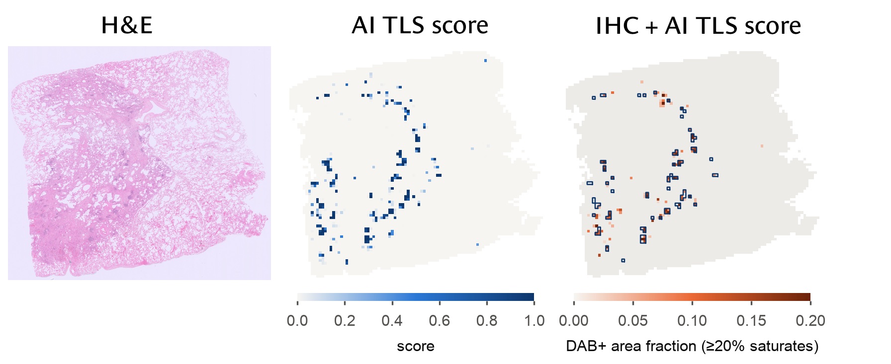
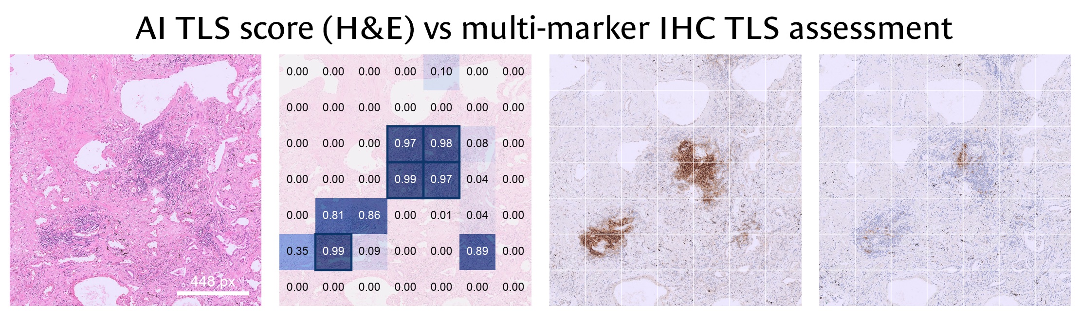
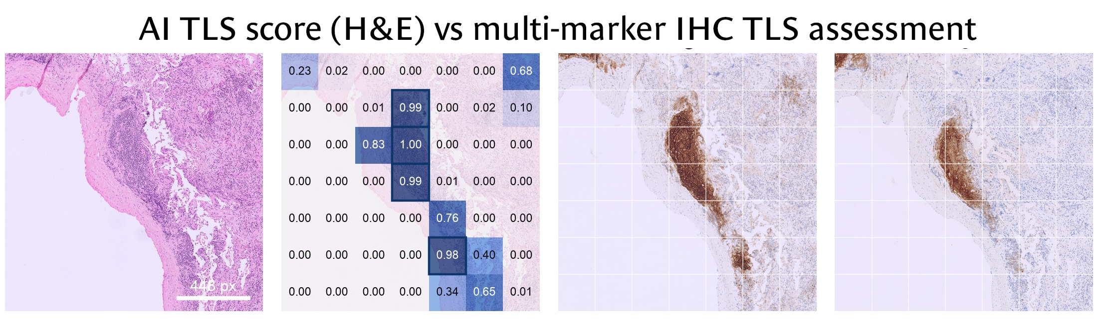
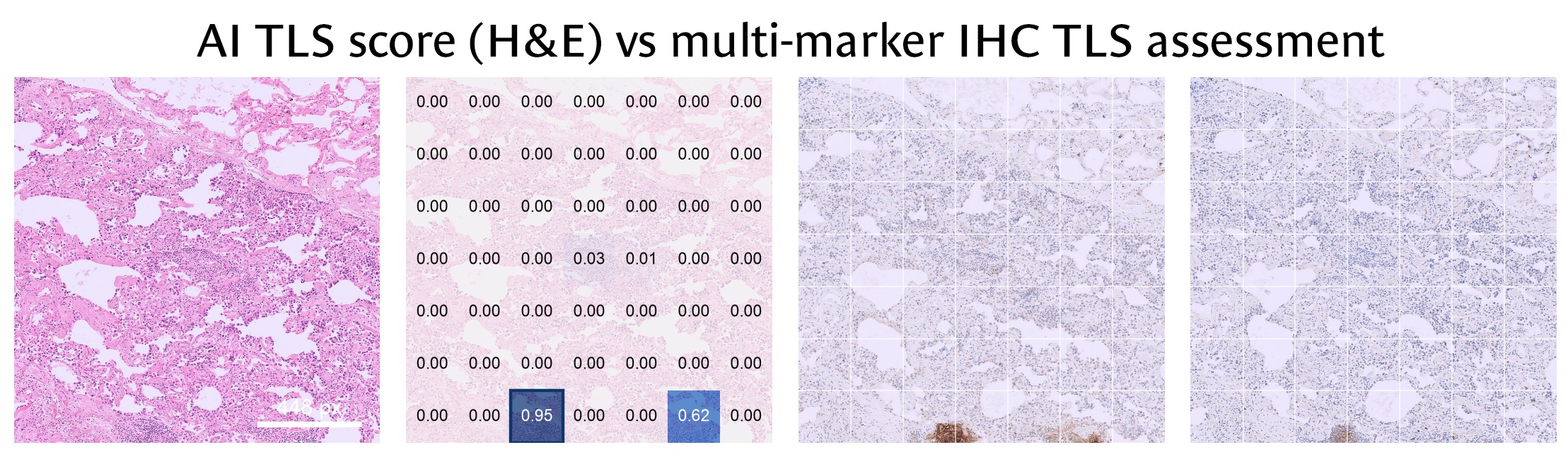
Where the model scores high on H&E, immunohistochemistry confirms TLS

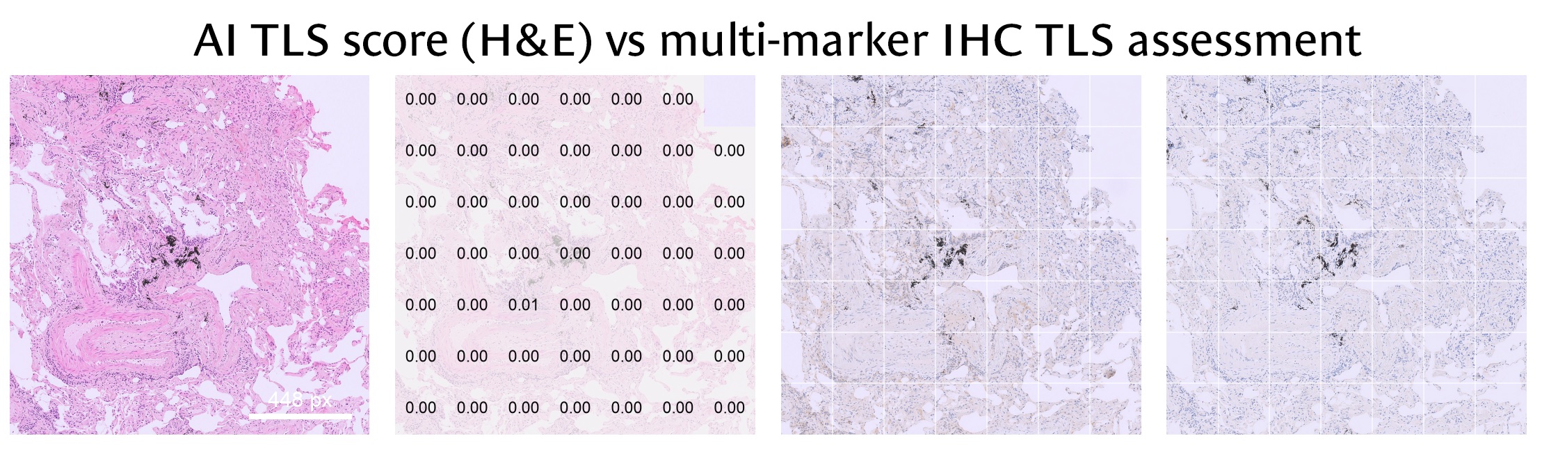
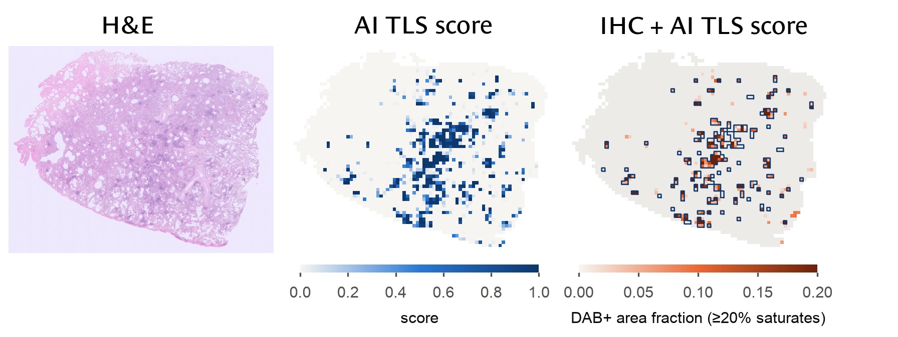
We compared the AI's TLS score on H&E with a multi-marker IHC assessment of TLS on slides aligned to the same tissue. The model never saw the IHC. The same check works with any IHC marker or panel, including your own.

Left to right: H&E · AI TLS score per tile · IHC staining (orange) with the AI's TLS tiles outlined

Whole lung slide: H&E, the AI TLS score per tile, and the IHC staining with the AI's TLS tiles outlined; the outlined tiles sit on stained areas
Slide 1. The tiles the AI scores as TLS cluster where the IHC stains.
Second whole lung slide: H&E, the AI TLS score per tile, and the IHC staining with the AI's TLS tiles outlined; the outlined tiles follow the stained areas
Slide 2. The same pattern on a slide with fewer, smaller TLS.
Explainability

Why the model identifies a region as TLS

The model's decisions align with features a pathologist can verify: dense, small nuclei, and a pattern that holds in every case. Hover over or tap the charts for values.

Tissue signature

How TLS differ from the surrounding tissue

Median inside annotated TLS divided by the median in the surrounding tissue. Values right of 1 are higher in TLS.

Consistency

Denser than the surrounding tissue in every image

Median nuclear density inside TLS and in the surrounding tissue of each image, sorted by the difference.

Inside TLSSurrounding tissue
Clinical survey · about 3 minutes

For pathologists, oncologists and researchers

Tell us where predicting immunotherapy response is hardest in your work, and whether the TLS analysis on this page would help. Your answers guide what we build through the ICURe Discovery programme; only your role is required.

Work in pharma, biotech, a CRO or a digital pathology company? Take the industry survey instead (about 8 minutes) →

What you saw on this page
Value of AI response prediction from H&E, compared with current biomarkers such as PD-L1
Credibility of the results on this page
Clarity of the AI's reasoning
Which elements were most useful? Select all that apply.
Where would you apply it? Select all that apply.
Next step
Would you like any of these? Select all that apply.
Contact & partnerships

Work with us

We are partnering with early adopters: pathology groups, translational researchers and industry teams who want TLS measurements from their own H&E slides.

  1. Tell us about your interest or question.
  2. We reply by email to arrange a short call.
  3. Together we scope a pilot on a sample of your slides.

Reason for contacting us * Select all that apply.